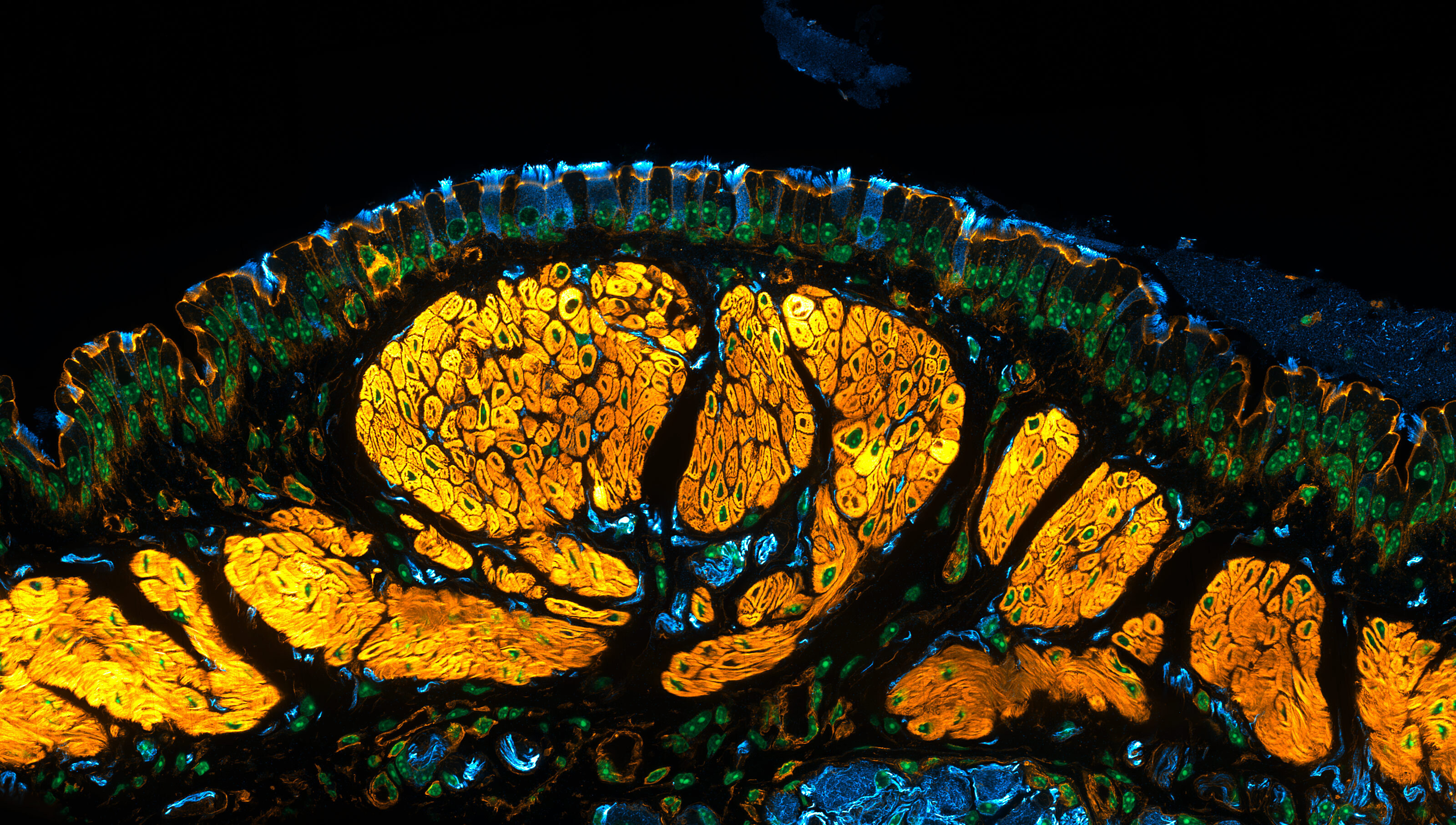
Since 1975, Nikon’s Small World competition has featured images produced by optical microscopes, an art that usually stays in scientific papers and textbooks. But through the carefully engineered lenses of the microscope, photomicrographers sometimes capture glimpses of strikingly surreal, alien beauty. The 2026 Photomicrography Competition winners include subjects both living and mechanical, small and very small.
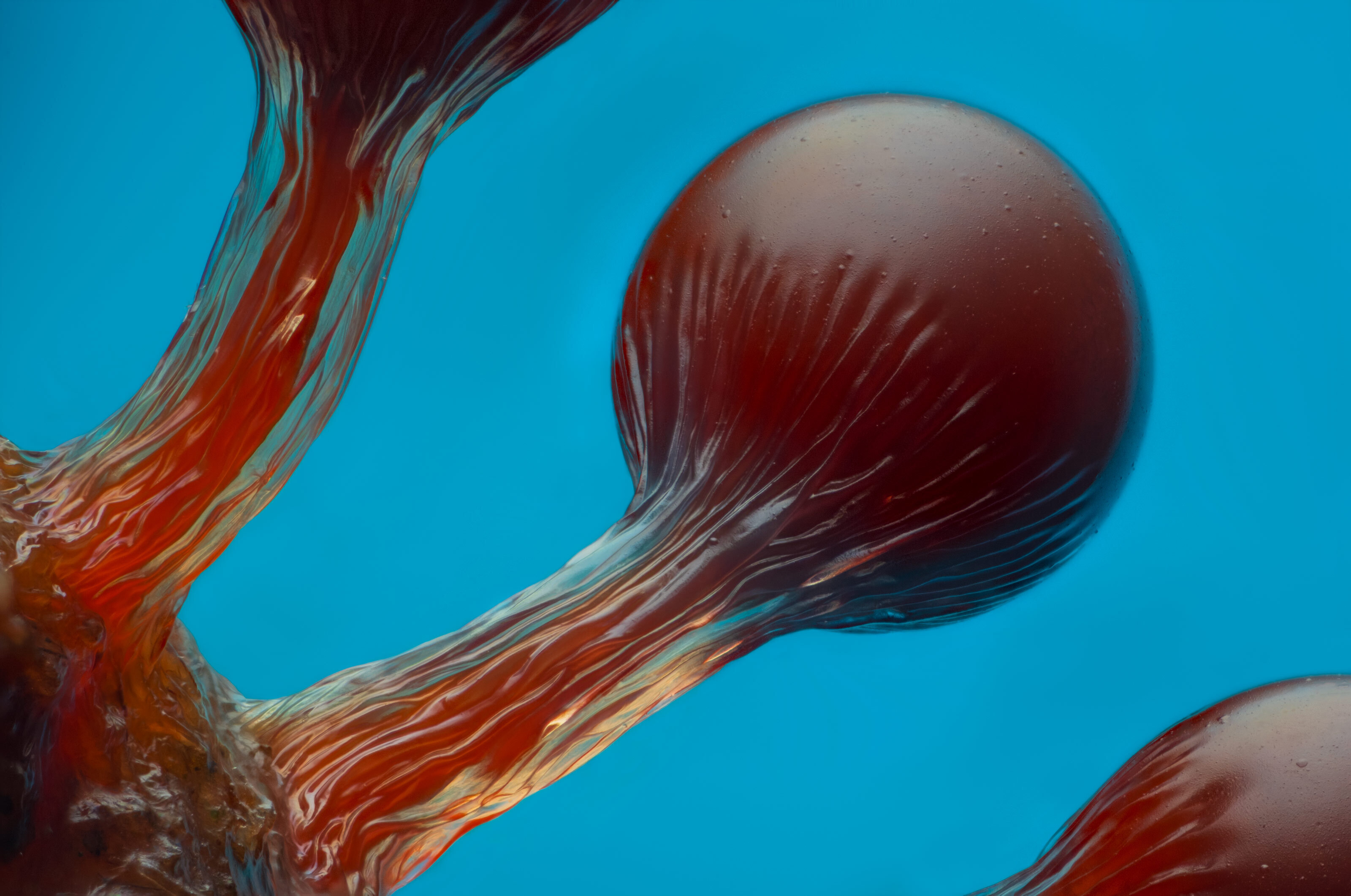
The fruiting bodies of a Hemitrichia slime mold remind one distinctly of Dr. Seuss. Photo: Eric Epstein/Nikon Small World
Inside the printer
The winning image shows bubbling globules of black ink being injected onto a piece of paper by an inkjet printer. Behind the lens was 25-year-old German design student Ole Bielfeldt, whose work explores the surprising complexity of everyday objects. His process is largely DIY: Bielfeldt builds his own setups, cobbles together parts, and learns techniques online.
“I just pick somewhere to start and go for it,” Bielfeldt explained. “You’re likely going to fail at first, and that’s what makes it even more interesting.”
The experimentation and imperfection, he added, are the most interesting parts of the process and, ultimately, more important to him than achieving the highest image quality.
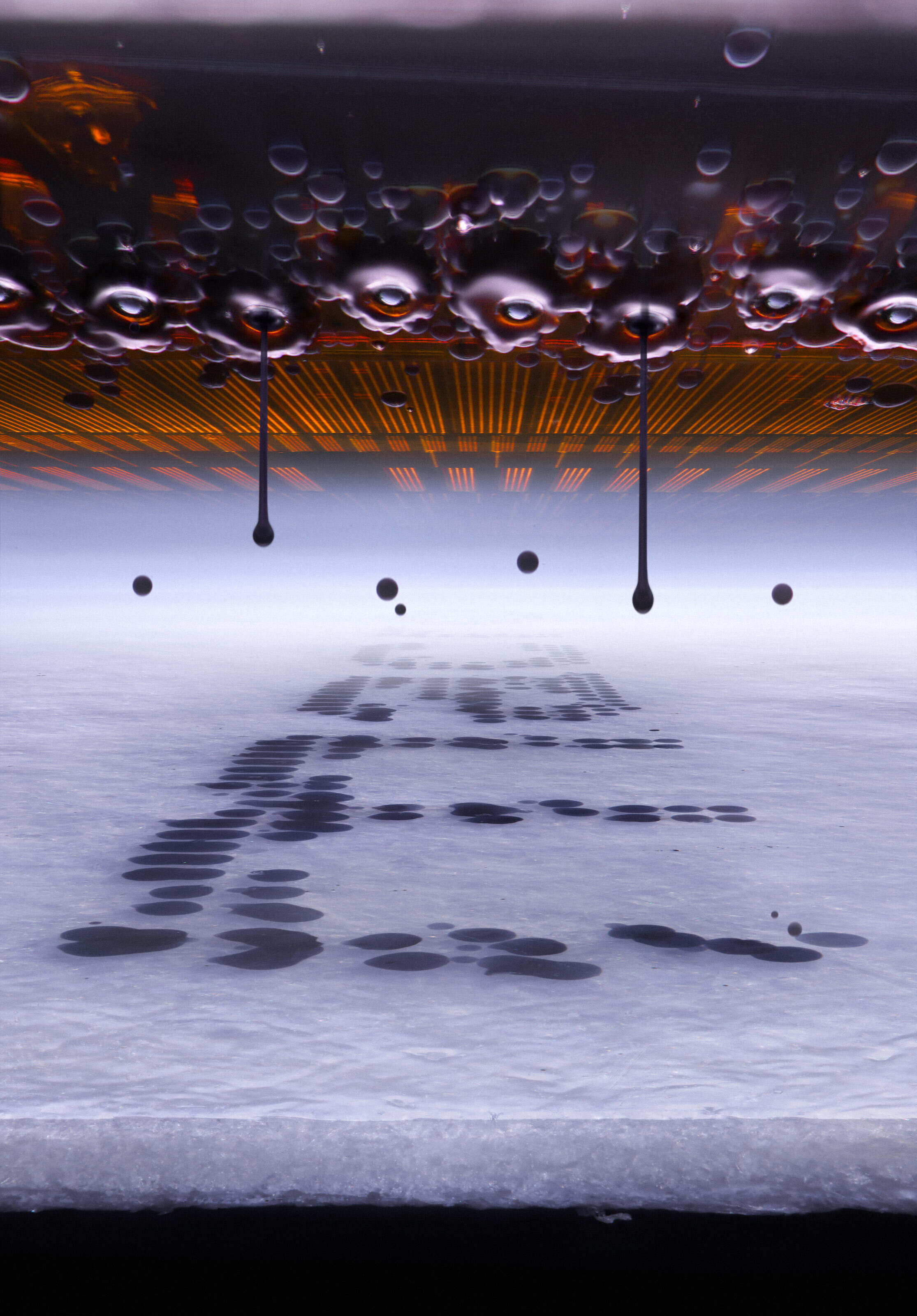
Photo: Ole Bielfeldt/Nikon Small World
Mouse parts surprisingly beautiful
Ah, mice; the humble martyrs to scientific progress. A significant amount of science seems to involve cutting up mice and learning from the result. But in this sort of photography, even the strongest mouse advocate must admit that the results are visually spectacular.
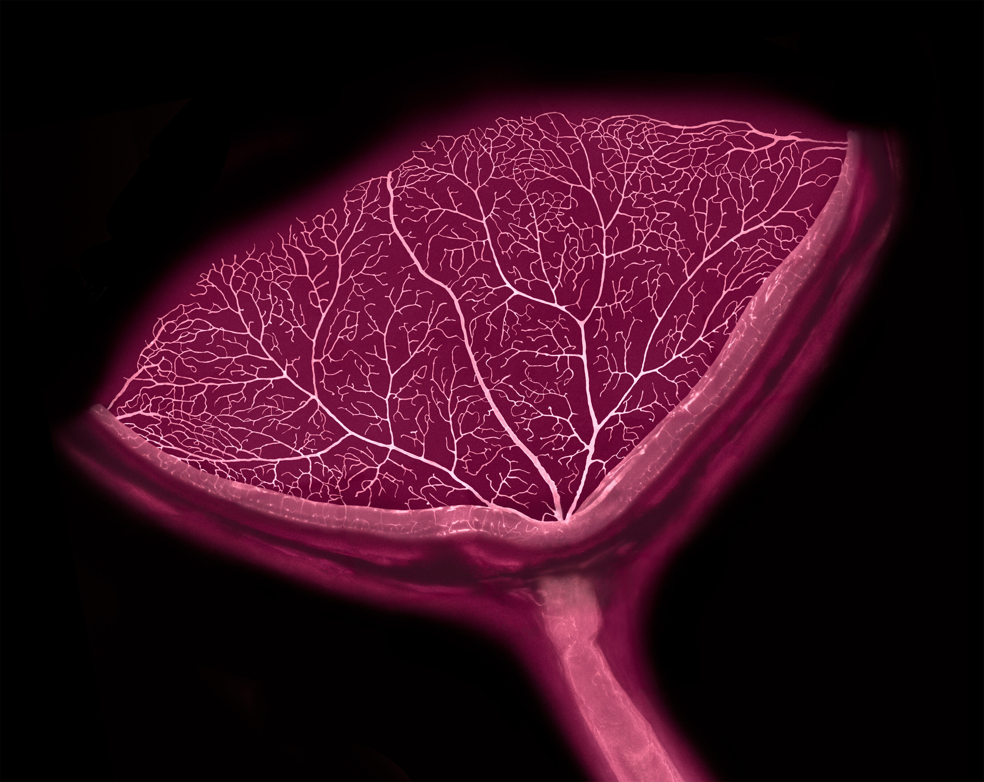
Satu Paavonsalo and Dr. Sinem Karaman of the University of Helsinki took this image of a mouse’s eye while researching drug therapy. Photo: Satu Paavonsalo and Dr. Sinem Karaman/Nikon Small World
Third place went to another rodent part: a 5X-magnified image of neurons in a mouse’s brain.
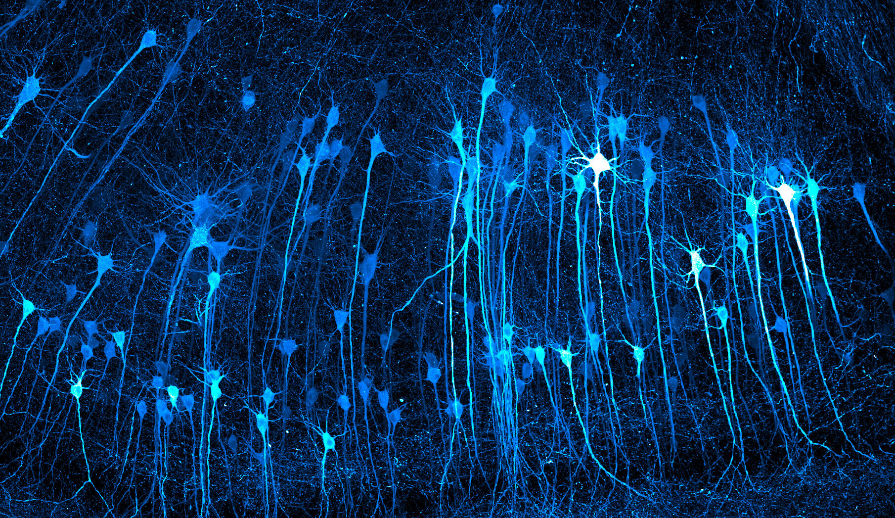
Photo: Kaelin Wulf and Rachel Flannery/Nikon Small World
In defense of humanity, we were also willing to subject our own body parts to photographic investigation. The 7th-place winner came from Dr. Bruno Cisterna and Dr. Eric Vitriol at the Medical College of Georgia at Augusta University. Using confocal imaging, which involves scanning a specimen to generate optical sections, they captured human neurons at 20X magnification.
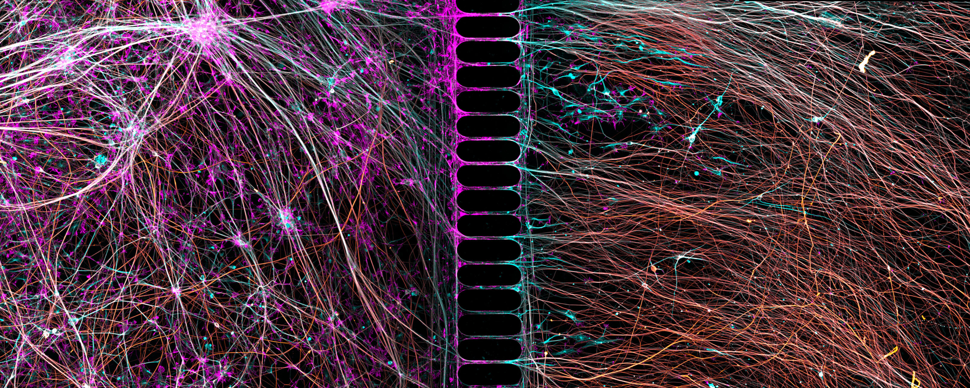
These human neurons were grown from reprogrammed skin cells. Photo: Dr. Bruno Cisterna and Dr. Eric Vitriol/Nikon Small World
I’ll end with a personal favorite of mine, John-Oliver Dum’s image of a little jumping spider, dusted with pollen. Dum, who won second place, credits a childhood bite for his fascination with spiders. The pollen was cultivated by his wife, specifically to create images like this one.
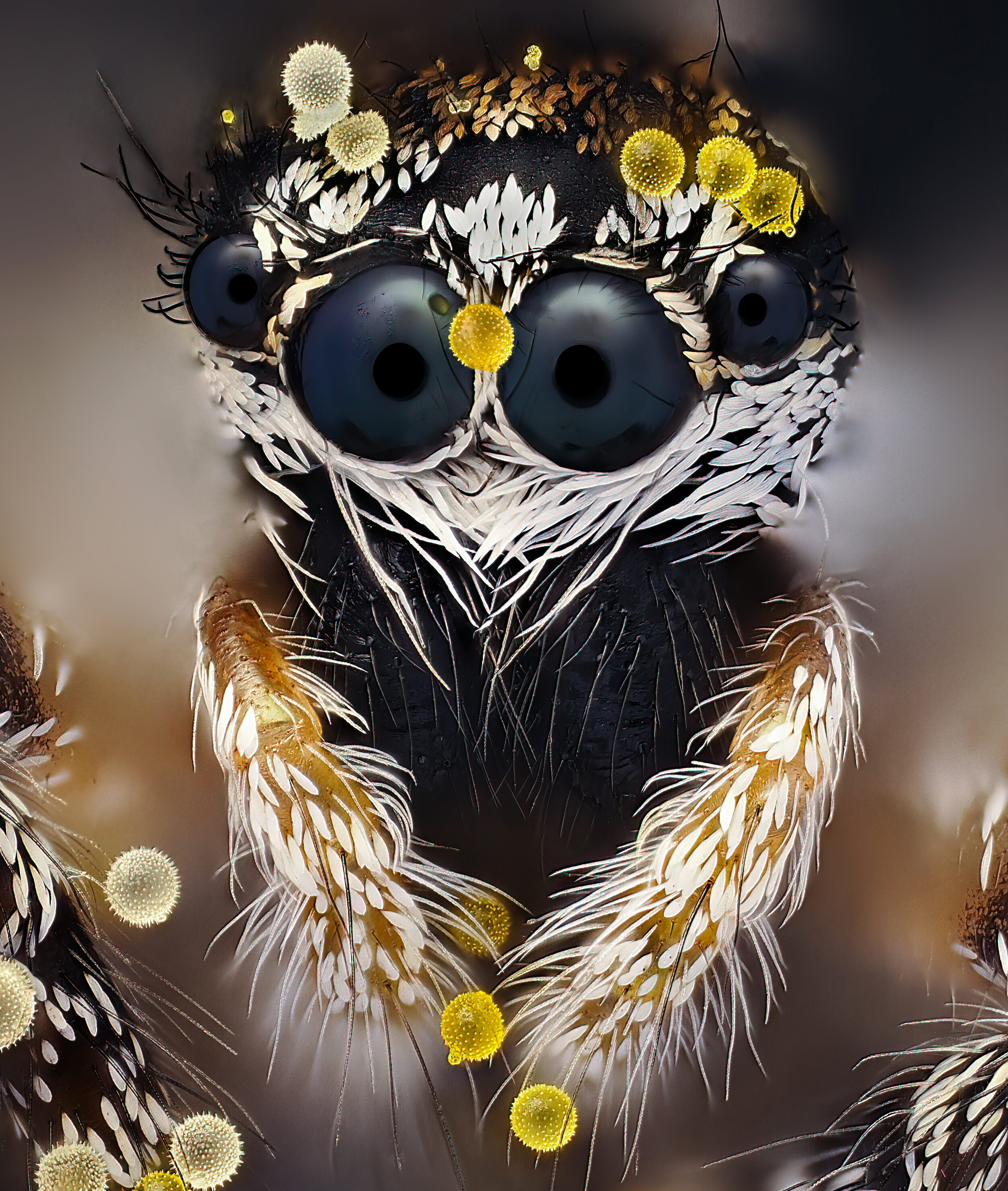
You can disagree, but I think this little guy is adorable. Photo: John-Oliver Dum
You can view the full gallery of winners and honorable mentions here. If you want to see photos of larger things, check out the winners from the Wildlife Photographer of the Year 2026, Aerial Photography, 2026 Ocean Photographer of the Year, or Memorial Maria Luisa International Photo and Video competitions.